
The Role of Imaging in Minimally Invasive Surgery
20-10-2026
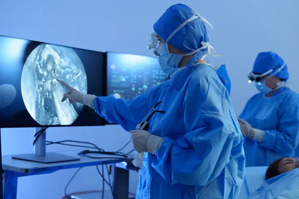
Seeing beyond the surgical field
Minimally invasive surgery has transformed how many procedures are performed. Smaller incisions, reduced tissue disruption and increasingly sophisticated instruments have opened the way to procedures that can offer patients less invasive treatment and, in appropriate cases, faster recovery.
But minimally invasive surgery also creates a fundamental challenge: the smaller the access point, the more limited the surgeon's direct view of the anatomy.
This is where imaging becomes more than a diagnostic tool. Increasingly, imaging is becoming part of the surgical process itself, helping clinicians locate anatomy, identify structures, guide instruments and make decisions while a procedure is underway.
Among the technologies supporting this evolution, ultrasound has a particularly important role because it can provide dynamic, real-time imaging without ionising radiation. Research reviews have identified intraoperative ultrasound as a valuable adjunct for lesion localisation, surgical guidance, vascular assessment and image-guided interventions.
From seeing the surface to understanding what lies beneath
Conventional endoscopic imaging provides surgeons with a direct view of the operative field. It is highly valuable, but it primarily shows surfaces.
Ultrasound can provide a different layer of information.
By generating images from within the body, ultrasound can help visualise soft tissue structures that are not directly visible through an endoscope or surgical access point. During surgery, this can support localisation of lesions, assessment of surgical margins and evaluation of blood vessels and tissue perfusion.
This distinction becomes particularly important when anatomy changes during a procedure. Organs can move, tissue can deform and structures can appear differently from their pre-operative position.
Pre-operative CT or MRI therefore cannot always provide the complete picture at the moment a surgical decision has to be made.
Intraoperative imaging can help bridge that gap.
A 2026 position paper from the German Society for Ultrasound in Medicine describes intraoperative ultrasound as a tool for surgical decision-making, including orientation, resection guidance, navigation in anatomically unclear situations and assessment of vascular anastomoses and organ perfusion.
Why real-time imaging matters
The value of intraoperative imaging is not simply the ability to produce another image. It is the ability to provide information when that information can influence the next surgical action.
In minimally invasive procedures, this can mean:
-
locating a target that is not visible from the surgical field
-
assessing the relationship between a lesion and surrounding anatomy
-
helping guide an instrument or therapeutic device
-
monitoring tissue during an intervention
-
evaluating blood flow or perfusion
-
identifying anatomical changes during a procedure
-
supporting decisions about the extent or direction of treatment
The distinction is important. Imaging is increasingly moving from being something clinicians consult before or after an intervention to something that can participate in the intervention itself.
The importance of miniaturisation
For imaging technology to become useful within minimally invasive procedures, the imaging system must fit within the constraints of the procedure.
That creates a demanding engineering problem.
A transducer may need to be small enough to pass through a catheter or other access route while still producing clinically useful images. It may also need to withstand repeated use, provide controlled steering and integrate with existing imaging platforms.
This is one of the areas in which Oldelft Ultrasound has developed specialist expertise.
Oldelft Ultrasound, based in Delft in the Netherlands, began developing ultrasound transducers in the 1980s and introduced its first transesophageal echocardiography (TEE) transducers in 1988. Today, the company develops 2D and 4D ultrasound transducers, with a particular focus on miniaturisation and tailor-made OEM solutions. Oldelft Ultrasound became part of steute Technologies in 2026.
Its technology illustrates the engineering direction of travel particularly well.
Oldelft Ultrasound's 4D ultrasound portfolio includes intracardiac echocardiography (ICE) technology designed for catheter-based imaging. Its 4D ICE transducer is designed to fit into a 12 French catheter and uses a 1,024-element acoustic array.
The significance is not simply that the probe is smaller. Miniaturisation can enable imaging to move closer to, or directly into the procedural environment.
From 2D images towards 3D and 4D information
Two-dimensional ultrasound remains an important imaging modality, but minimally invasive and robotic procedures increasingly demand more spatial information.
Three-dimensional and 4D ultrasound can provide volumetric information in real time, potentially helping clinicians understand anatomy and instrument position in three dimensions rather than relying on a sequence of two-dimensional views.
Research into ultrasound guidance for minimally invasive robotic procedures has highlighted real-time volumetric imaging as one of ultrasound's advantages and identified its potential to help clinicians visualise changing soft-tissue anatomy during procedures.
The technology is also evolving beyond simply producing a better image.
Advanced ultrasound techniques, image reconstruction, spatial tracking and multimodal image fusion are being investigated to support increasingly sophisticated image-guided procedures.
This points towards an operating environment in which imaging, surgical instruments, navigation systems and robotics increasingly work as interconnected components rather than separate technologies.
Imaging where the intervention happens
Perhaps the most significant development is the movement of imaging closer to the point of intervention.
“Oldelft Ultrasound's work in the SEISMIC-IHI research programme provides a useful example. The five-year, €23.5 million programme aims to advance minimally invasive treatment of brain tumours, with Oldelft Ultrasound developing a 4D ultrasound probe intended to visualise the brain during cryoablation. The objective is real-time imaging guidance within the clinical procedure.
This illustrates a broader principle: the future of minimally invasive surgery depends not only on smaller instruments, but also on smaller and more capable imaging technologies.
The better imaging can can visualize the treatment site, the more directly it can inform the procedure”, says Michiel Baas, R&D Manager at Oldelft Ultrasound, in Delft, The Netherlands.
The engineering challenge behind the image
Producing a clinically useful image from a highly miniaturised transducer is not straightforward. It requires advances in acoustic design, electronics, signal processing, materials, mechanical construction and system integration.
Oldelft Ultrasound's 4D platform, for example, uses an in-house ASIC architecture and matrix-array technology; scalable across different 4D applications, including catheter-based imaging and miniaturised TEE solutions.
The engineering challenge is therefore not simply to reduce the size of an existing probe. It is to preserve, and where possible, enhance imaging capability while reducing the physical footprint.
That requires close collaboration between transducer engineers, medical device manufacturers, clinicians and imaging-system developers.
The role of OEM collaboration
A transducer is part of a larger system involving imaging hardware, software, displays, navigation, surgical instruments and clinical workflows. For this reason, the development of imaging technology increasingly depends on collaboration between specialist technology companies and medical device OEMs.
Oldelft Ultrasound describes its business model around long-term OEM partnerships and tailor-made transducer development, including applications in cardiology, surgery and urology.
For medical device manufacturers, this type of specialist expertise can be particularly valuable when an application requires imaging technology that cannot simply be adapted from a conventional diagnostic probe.
Where imaging and minimally invasive surgery are heading
The direction of development is clear, although many technologies remain at different stages of clinical validation and adoption.
Future minimally invasive procedures are likely to involve increasingly sophisticated combinations of:
-
miniaturised ultrasound transducers
-
3D and 4D imaging
-
robotic surgical systems
-
image-guided navigation
-
artificial intelligence and image analysis
-
multimodal imaging
-
real-time procedural data
The objective is not technology for its own sake, it is to provide clinicians with relevant information at the point when a decision needs to be made.
Intraoperative ultrasound is already used across several surgical specialties, but its integration into routine surgical practice remains uneven. Recent clinical literature has called for more structured integration of intraoperative ultrasound into training, guidelines and surgical workflows.
That suggests the next stage of progress will depend not only on improving image quality, but also on making advanced imaging practical, interoperable and clinically meaningful.
From imaging technology to procedural intelligence
Minimally invasive surgery began by changing how surgeons access the body.
The next phase is changing how they see and understand it while they operate.
As transducers become smaller, imaging becomes more three-dimensional and systems become increasingly integrated, the boundary between imaging and intervention is becoming less distinct.
“The future operating environment may not be defined simply by smaller instruments or smaller incisions. It may be defined by the ability to combine precise intervention with continuous, actionable information about the anatomy being treated. That is where technologies such as miniaturised 2D and 4D ultrasound can play an important role.
For companies developing the next generation of medical devices, the challenge is therefore not simply to create an imaging probe. It is to engineer the interface between imaging, intervention and clinical decision-making. And increasingly, that interface needs to be small enough to go where the procedure goes.” says Erwin Dumay, Managing Director at Oldelft Ultrasound.
About Oldelft Ultrasound
Oldelft Ultrasound is a medical diagnostic company, specialized in the development, manufacture and service of innovative Ultrasound transducers. Oldelft Ultrasound is an OEM supplier to the Ultrasound equipment companies worldwide. Oldelft Ultrasound is especially well-known for its TEE (Trans Esophageal Echocardiography) transducers, and specialists in miniaturization of transducers. Based out of Delft, in The Netherlands, Oldelft Ultrasound is part of the steute Technologies group.
Oldelft Ultrasound - Commercial Contact
Stefan Roggeveen info@oldelft.nl